Lay out labeled leads and plug them into their designated outlets on the 15-lead electronics box. V4 At the mid-clavicular line in the fifth intercostal space.

Ecg Educator Blog Posterior Ecg Lead Placement
ECG Monitoring 1215 Lead PlacementResources.

. Lead Placement for Posterior ECG. Total scene time should not exceed 20 minutes. ECG Lead Placement and Identifying Lead Reversal This quick reference guide is intended to show correct ECG electrode locations and how to recognize inadvertent lead wire reversal.
Continuing Medical Education Section 1. Right sided 12 lead ECG lead placement. ECG Monitoring 12 -Lead.
Basic 12-Lead Placement 1. NOTELead placement may vary by institution or instruction. Posterior extension of an inferior or lateral infarct implies a much larger area of myocardial damage with an increased risk of left ventricular dysfunction and death.
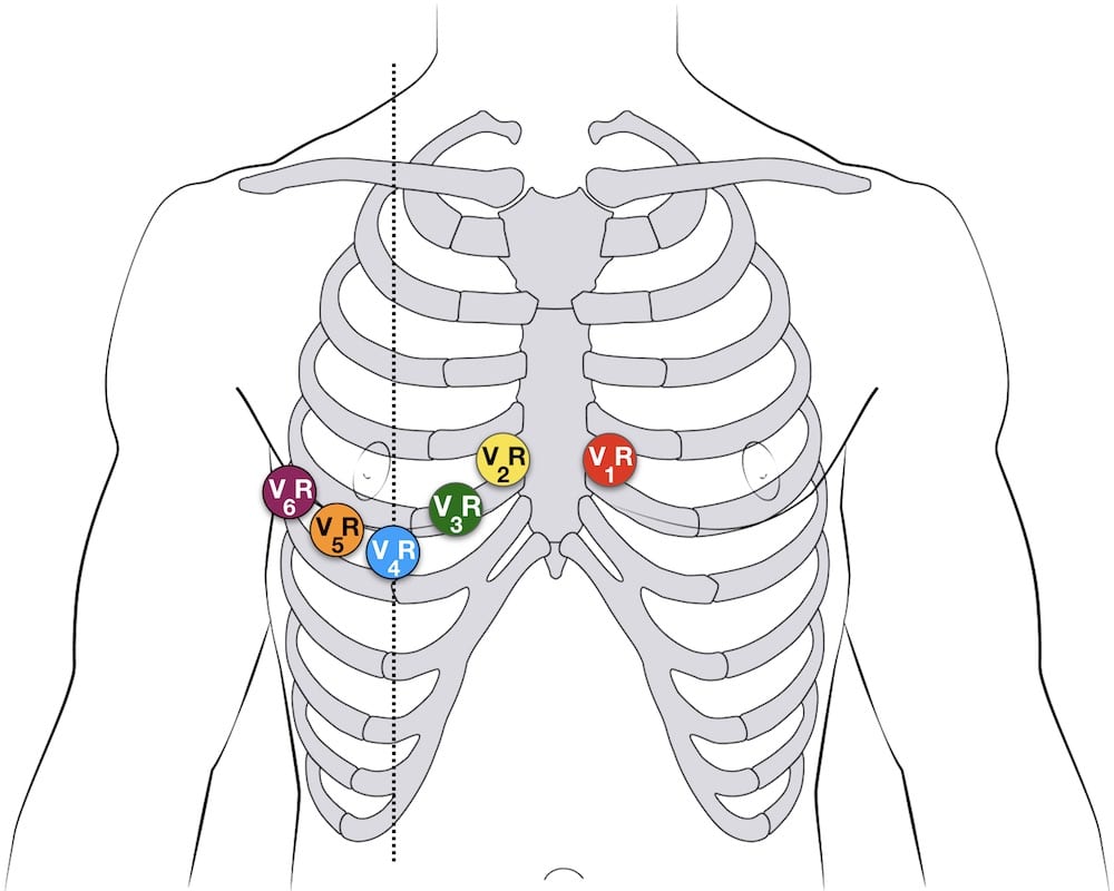
Lead Electrode Placement15 lead Preparation and Placement V1 Fourth intercostal space next to the sternum on the right side. 12-Lead ECG Interpretation Introduction This self-study package has been developed to provide a review of twelve lead interpretation as well as a review of signs and symptoms of various types of AMIs. Limb lead placement For accurate 12-lead measurements and interpretation limb leads must be placed on the limbs not the torso.
The leads V4-V6 are removed and substituted for V7-V9 as shown below. Basic 12-Lead Placement 1. Leads V7-V9 was 26.
To detect posterior infarcts which are often associated with inferior or lateral wall AMI. While the 18-lead ECG is perhaps more sensitive for early detection of ischemia or infarction in practice either should be used for. They are performed by placing V4 V5 and V6 electrodes in the same intercostal space but continuing into the patients back.
Besides the incidence of isolated posterior MI is not defined and has been reported in studies ranging from 0 to 7-12 18 23. Isolated posterior MI is less common 3-11 of infarcts. Enter the patients name and date of birth for all 12- leads day 2 month 3 year 4 on the cardiac monitor if the day is a single digit do not preface with.
RS amplitude ratio in V1 or V2 is 1. It can be simpler to leave V1 and V2 in their usual positions and just transfer leads V3-6 to the right side of the chest ie. FWIW heres the placement theory and rationale shes using.
See figures 8 9 3. Lead ECG taken from 50 IWMI patient s the overall incidence of ST elevation in the posterior chest. On most EKg machines the labels areno automatically changed so it is important to cross out the labels for V4-V6 and write in V7-V9.
ECG chest precordial lead placement left anterior oblique view Finding the Correct Placement of Leads V1 V6. 12- 15- lead ECG Section 1. Ill do a right 15 or 18 lead if Im really suspicious of something cardiac going on but cant immediately find it on a 12 lead or if I see an inferior wall MI.
When viewing the EKG strip V4-V6 on the strip will be referred to as V-13-15. 2 patients among the 50 had both RVI and PWMI. 4-5 Indications of a posterior wall infarction may include.
V2 Fourth intercostal space next to the sternum on the left side. Posterior Leads Electrodes Placement for Posterior Leads Posterior leads are helpful in suspected posterior myocardial infarction. V4V7 V5V8 and V6V9.
In the fifth intercostal space and the left posterior axillary line. A prehospital 12-lead ECG may be initiated and performed on scene but should not extend scene time. In this series of 15 -.
Nasco Life form 15 - Lead ECG Placement Trainer teaches up to 15 - Lead ECG electrode Placement anatomically and provides visual feedback on accuracy of electrode placement. So far weve identified one posterior STEMI that wouldnt have shown on a 12 weve been running 15s since last August-ish. When viewing the EKG strip V4-V6 on the strip will be referred to as V-13-15.
When a 15-lead or 18-lead ECG machine is not available manipulation of the leads from a standard 12-lead ECG machine allow additional areas of the heart to be imaged. 15 and 18 Lead ECG. 12- 15- lead ECG Section 1.
A standard pre-hospital 15 lead ECG consists of taking a second 12 lead ECG but moving 3 electrodes to V4R V8 and V9. Hints of an associated posterior infarct. Aside from a 12-lead ECG placement theres something known as a 15-lead placement which includes placing leads V4-V6 on the posterior side of the patient below their left scapula see below.
Ensure the trainer is clean. Doing a 15 lead ECG Firstly do a standard ECG then by repositioning leads V4 V5 and V6 to the patients back they become V7 V8 and V9. It is also helpful for future clinicians if you note in your read that it is a posterior ECG.
15 or 18 lead ECGs can be done with alternate precordial lead placement to assess for posterior- or right-sided disease. V3 Midway between locations V2 and V4. In addition the use of the 15-lead ECG confirms the posterior MI and is superior to the findings in the anterior leads.
Locate the sternal notch Angle of Louis by feeling the top portion of the breast bone and moving your fingers downward until you feel a bump. Posterior infarction accompanies 15-20 of STEMIs usually occurring in the context of an inferior or lateral infarction. Aside from a 12-lead ECG placement theres something known as a 15-lead placement which includes placing leads V4-V6 on the posterior side of the patient below their left scapula see below.
A complete set of right-sided leads is obtained by placing leads V1-6 in a mirror-image position on the right side of the chest see diagram below. Posterior ECG leads V7-V9 are applied by moving V4-V6 to under the left scapula. When viewing the EKG strip V4-V6 on the strip will be referred to as V-13-15.
Placement of Lead V1. STD in V1-V3 or. The last time I did a posterior EKG was on a guy who told me he last had a posterior wall MI.
Where do you place a 15 lead ECG. Aside from a 12-lead ECG placement theres something known as a 15-lead placement which includes placing leads V4-V6 on the posterior side of the patient below their left scapulasee below. Posterior infarction accompanies 15-20 of STEMIs usually occurring in the context of an inferior or lateral infarction.
Lead Placement for Posterior ECG Resus Review. To clarify leads will equal.

Aliem Cards

Ppt Chapter 8 For 12 Lead Training The 15 Lead Ecg Powerpoint Presentation Id 294278
Placement Of Posterior Chest Leads For Identifying Left Circum Ex Download Scientific Diagram

Ecg Lead Positioning Litfl Ecg Library Basics

All Posterior Positioning Of The Electrocardiographic Leads On The Download Scientific Diagram
Ecg Lead Positioning Litfl Ecg Library Basics

Aliem Cards

Importance Of Posterior Chest Leads In Patients With Suspected Myocardial Infarction But Nondiagnostic Routine 12 Lead Electrocardiogram Sciencedirect
0 comments
Post a Comment